
3D MEA
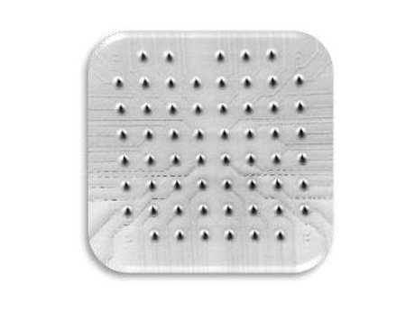



Reliably Record Electrical Activity from Inside Tissue Slices and Organoids
Traditional planar 2D microelectrode arrays record activity at the tissue surface. 3D MEAs extend beyond the surface using penetrating three-dimensional microelectrode array technology, enabling reliable access to electrical activity within thick tissues and 3D cell cultures by placing recording sites closer to active tissues.
Featuring a unique, precision-engineered spike electrode design, 3D MEAs are designed to record from viable cell populations located below the tissue surface, providing a powerful solution for studies involving acute hippocampal slices, organotypic slices, brain organoids, retinal organoids, and other advanced tissue models.

What Is a 3D Multielectrode Array?
3D MEAs incorporate elevated penetrating electrodes rather than conventional planar recording sites. These elevated electrodes (also referred to as spike electrodes or conical electrodes) extend into the tissue to access active cells located beneath the sample surface, making them ideal for applications where neuronal cell bodies are distributed throughout a three-dimensional structure or where superficial cell layers may not provide the strongest electrophysiological signals.
The result is improved access to neuronal activity within thicker tissues while maintaining the advantages of multi-electrode, network-level recording.
Watch a Webinar on 3D MEA and Organoid Research (link is external) See Publications


3D MEA for Acute Tissue Slices
3D MEAs are widely used for electrophysiological recordings from acute neural tissue preparations, including:
- Acute hippocampal slices
- Cortical slices
- Cerebellar slices
- Organotypic slices
- Retinal explants
For acute slice experiments, recordings are often enhanced by positioning electrodes closer to healthy neuronal populations located beneath the cut surface. Following tissue preparation, superficial layers can contain damaged or less active cells, while deeper regions may retain stronger physiological activity. Penetrating electrodes provide access to these regions without the need for multiple individually positioned needle electrodes.
What Are the Advantages of 3D MEA for Slice Electrophysiology?
- Access to neuronal activity below the tissue surface
- High spatial resolution network recordings
- Simultaneous recordings across large tissue regions
- Compatibility with live-cell imaging
- Streamlined workflows compared with single-probe approaches
3D MEAs are particularly valuable for researchers seeking detailed network activity from acute brain slices while preserving the spatial context of the tissue.
Contact Us to Learn More Shop 3D MEAs Online (link is external)

3D MEA for Organoids and 3D Cell Cultures
Organoids present unique recording challenges due to their inherently three-dimensional architecture. In many models, electrically active cells are distributed throughout the tissue volume, making it difficult to predict where the strongest signals will originate. For these applications, recording from inner cell layers is often essential.
3D MEAs can record from within
- Brain organoids
- Cortical organoids
- Neural spheroids
- Retinal organoids
- Cardiac organoids

What Are the Advantages of 3D MEA for Organoid Research?
- Access to neurons distributed throughout the organoid
- Recording from deeper tissue regions
- Longitudinal monitoring of network development
- Compatibility with established submerged culture workflows
- High spatial resolution electrophysiological measurements
Watch a Webinar on 3D MEA and Organoid Research (link is external) Schedule a Meeting with Our Team (link is external)
3D MEA, Mesh MEA™, or ALI MEA™ for Organoid Electrophysiology?
As organoid models become increasingly complex, researchers need tools that support long-term culture while still providing reliable access to electrophysiological data. While 3D MEAs continue to be a proven solution for recording from complex 3D tissues, researchers can now also choose from newer platforms optimized for specific workflows, including long-term organoid culture and air-liquid interface applications.
Depending on experimental goals, Mesh MEA™ or ALI MEA™ may offer additional advantages over traditional 3D MEA approaches.

Mesh MEA™ enables long-term electrophysiological recordings from the interior of intact 3D tissue models. By allowing cells to grow naturally around the mesh and electrodes, Mesh MEA preserves physiologically relevant morphology while providing access to functional signals from throughout the tissue structure.

ALI MEA™ integrates electrophysiology, perfusion, and air-liquid interface culture in a single platform to support healthier tissues, simplified workflows, and more reproducible recordings. Better oxygen supply due to air-liquid interface. ALI MEA helps researchers generate high-quality data while reducing preparation and handling complexity
Learn More About ALI MEA More Organoid Research Solutions from HBio (link is external)
Multiple Electrode Density and Spacing Options
The optimal electrode layout depends on the size of the biological sample and the desired recording resolution. Larger tissues typically benefit from greater recording area coverage, while smaller preparations often benefit from increased electrode density.
Higher Density Arrays
Ideal for:
- Small organoids
- Local network analysis
- Fine spatial mapping
Wider Electrode Spacing
Ideal for:
- Large tissue slices
- Human tissue preparations
- Broad network coverage

| Product name | Compatible systems | Interelectrode distance | Electrode diameter | Electrode height | |
|---|---|---|---|---|---|
| 60-3DMEA100/12/40iR-Ti | Shop Online | 100 µm | 12 µm | 40 µm | |
| 60-3DMEA200/12/50iR-Ti | Shop Online | MEA2100-Mini-60 AcadeMEA™ 60 |
200µm | 12 µm | 50 µm |
| 60-3DMEA200/12/80iR-Ti | Shop Online | MEA2100-Mini-60 AcadeMEA™ 60 |
200µm | 12 µm | 80 µm |
| 60-3DMEA250/12/100iR-Ti | Shop Online | MEA2100-Mini-60 AcadeMEA™ 60 |
250µm | 12 µm | 100 µm |
| 120-3DMEA250/12/100iR-Ti | Shop Online | 250µm | 12 µm | 100 µm | |
Compatible Recording Systems
3D MEAs are compatible with the MEA2100-Mini System or AcadeMEA™ Single-Well MEA System.
Discover the Right 3D MEA Configuration
Contact the Multi Channel Systems team online or schedule a meeting with us to discuss your requirements and identify the best 3D MEA for your application.
Featured 3D MEA Publications
- De Rosa F, Kilb W, Luhmann HJ, Sinning A (2026) Network integration of neocortical Cajal-Retzius neurons shapes early cortical dynamics and contributes to their transient nature.
- Migliorelli C, Togninalli M, Scaparra A, Petrini EM, Ventre M, Chiappalone M (2026) Unraveling Hydra bioelectrical activity on multielectrode array.
- Nieto-Estevez V, Varma P, Mirsadeghi S, Caballero J, Gamero-Alameda S, Hosseini A, Silvosa MJ, Thodeson DM, Goswami S, Lybrand ZR, Giugliano M, Navara C, Hsieh J (2026) Dual developmental effects of ARX poly-alanine mutations on human cortical excitatory and inhibitory neurons.
- Meyer-Acosta KK, Diaz-Guerra E, Varma P, Aruk A, Mirsadeghi S, Muniz-Perez A, Rafati Y, Hosseini A, Nieto-Estevez V, Giugliano M, Navara C, Hsieh J (2025) APOE4 impacts cortical neurodevelopment and alters network formation in human brain organoids.
- Yin J, Lees JG, Gong S, Nguyen JT, Phang RRJ, Shi Q, Huang Y, Kong AM, Dyson JM, Lim SY, Cheng W (2025) Real-time electro-mechanical profiling of dynamically beating human cardiac organoids by coupling resistive skins with microelectrode arrays.
- Alberio L, Coulon P, Mosser S, Knöpfel T, Dallerac G (2024) MEA-NAP: MEA-NAP: A flexible network analysis pipeline for neuronal 2D and 3D organoid multielectrode recordings.
- Yadav N, Di Lisa D, Giacomozzi F, Cian A, Giubertoni D, Martinoia S, Lorenzelli L (2023) Development of multi-depth probing 3D microelectrode array to record electrophysiological activity within neural cultures.
- Lyu Q, Gong S, Lees JG, Yin J, Yap LW, Kong AM, Shi Q, Fu R, Zhu Q, Dyer A, Dyson JM, Lim SY, Cheng W (2022) A soft and ultrasensitive force sensing diaphragm for probing cardiac organoids instantaneously and wirelessly.
- Kwiatkowski CS, Shah S, Kylilis N, Tsaneva-Atanasova K, Murray AF, Melhuish C, Polizzi KM (2019) Formation of neural networks with structural and functional features consistent with small-world network topology on surface-grafted polymer particles.
Contact Us to Learn More Schedule a Meeting with Us (link is external)