You are here
Organoid research is breaking new ground. How will Mesh MEA transform it even further?

Founded nearly 30 years ago, Multi Channel Systems (MCS) is a pioneer in the development of commercially available microelectrode arrays (MEA) and their recording systems. MEAs have proven valuable for our understanding of how genetic background, disease pathology, and drugs alter neuronal network activity.

Organoids represent a unique experimental framework for studying human physiology and disease, but limitations in readout platforms create a barrier to the evolution of organoid research. MEA (multielectrode array) systems offer a way to record electrophysiological data from outside an organoid, but not without impacting its structure and thus compromising the validity of the data.
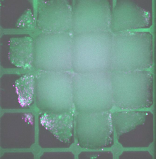
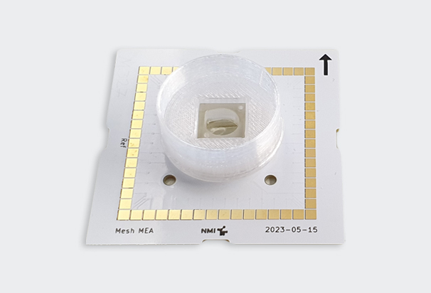
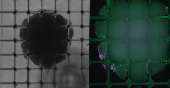
Mesh MEA from Multi Channel Systems is a revolutionary new technology that offers a number of benefits over traditional planar MEA chips, including 3D conformability, improved signal access, enhanced long-term recordings, and better representation on in vivo condition by adapting the organoid’s 3D shape.
1. Neural Organoid Studies in Neurodevelopment and Disease Modeling
Some research has used neural organoids to investigate abnormalities in neural networks, which may reveal differences in the brain cells of individuals with or without certain diseases or neurodevelopmental disorders. Using Mesh MEA to record electrophysiological data from inside the organoids, researchers could obtain more detailed, long-term recordings of electrical activity from different regions of the 3D organoids, enabling them to map neural circuitry abnormalities more precisely and observe how these networks evolve over time.
Core improvement with Mesh MEA: The ability to record from more sites within the organoid would provide deeper insights into how synaptic connections form, synchronize, and adapt – particularly in neurodevelopmental disorders (such as autism spectrum disorder), where early network dysfunction plays a key role.

2. Alzheimer's Disease Organoid Models
Brain organoids have been used to model Alzheimer’s disease by exposing them to amino acids that may lead to plaque formation and eventually neuronal death. In these models, understanding how electrical activity in neural networks is disrupted over time is crucial. Conventional MEA systems provide limited data on long-term changes in electrical connectivity.
Core improvement with Mesh MEA: Mesh MEA would allow for high-resolution tracking of neural firing patterns in different layers of the organoid, offering a more comprehensive view of how plaques affect network-level communication and neuron synchronization, potentially revealing subtle changes in network dynamics before major neuronal loss occurs.
3. Parkinson's Disease Modeling
Studies that generate dopaminergic neurons from stem cells and use them to model Parkinson’s disease typically focus on the progressive loss of these neurons. A key aspect of Parkinson's disease is the loss of coordinated electrical activity in the brain's motor networks. Traditional methods often miss the complex electrophysiological changes that occur during this degeneration.
Core improvement with Mesh MEA: Researchers could have used Mesh MEA to map the intricate electrical communication in Parkinson's organoids as dopaminergic neurons degenerate. This would improve understanding of how motor control-related neural networks degrade over time and how these networks respond to treatments like dopamine agonists or neuroprotective agents.
4. Cardiac Organoid Research
Cardiac organoids created from induced pluripotent stem cells (iPSCs) have been used to model heart diseases, including arrhythmias and congenital heart defects. Studies often involve electrophysiological assays to measure heart rhythm and detect irregularities, but traditional MEA systems have limitations in recording detailed electrical maps from the organoids.
Core improvement with Mesh MEA: Mesh MEA’s ability to record high-density, long-term electrophysiological data would provide a more precise mapping of how electrical signals propagate through the 3D structure of cardiac organoids. This would improve the detection of subtle changes in heart rhythms and provide better insights into arrhythmia mechanisms.
5. Organoid Studies for Drug Testing and Screening
Organoid models have been used to test the efficacy of chemotherapy drugs on cancerous tissues. Tracking how tumor cells respond electrically to drug treatments over time is crucial for understanding the drug's effects on cellular communication and growth.
Core improvement with Mesh MEA: The Mesh MEA system could capture real-time, long-term changes in tumor organoid electrophysiology in response to different drugs. This would allow for more dynamic monitoring of how treatments affect cancer cell behavior and network properties, potentially leading to more personalized and effective therapies.
6. Schizophrenia and Neural Dysfunction
Researchers have developed brain organoids from patients with schizophrenia to study neural connectivity and how it differs from healthy controls. One limitation in these studies is capturing the full complexity of dysfunctional electrical activity in these 3D models.
Core improvement with Mesh MEA: Using Mesh MEA could provide a more comprehensive picture of how neural activity becomes dysregulated in schizophrenia organoids. It could track the progression of network dysfunctions and offer insights into early intervention points for treating the disorder.
Conclusion
Mesh MEA offers improved signal access and the ability to record long-term electrical activity from multiple sites within 3D organoids. This would enhance the precision and depth of studies in neural network dynamics, disease progression, drug testing, and more. By capturing finer details of electrical communication across entire organoids, researchers could obtain more accurate data and develop better therapeutic strategies for diseases like Alzheimer's, Parkinson's, and various cardiovascular disorders.
